This article is the first in a 3-part series to help horse owners see and understand where their horse is lame.
Forelimb Lameness
As a veterinarian, I frequently encounter client questions as to how to evaluate lameness. In this article I’ll cover forelimb lameness and the common signs we look for, how to appraise the lame limb, and possible diagnostics a veterinarian uses once we arrive on the farm. Ultimately, any one of these topics could be a series of articles on their own, so if you have further questions feel free to contact me or your personal veterinarian.
Forelimb lameness is the obvious place to start. The front legs are generally the easiest of the limbs to diagnose, and problems can be seen in multiple ways. With ranges of lameness, the easiest to assess is when the horse is “pointing” or non-weightbearing. As lameness becomes more subtle, we look to other areas.
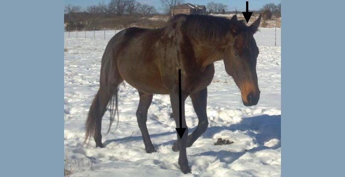
- The sound leg is the one the head and neck drops down on when moving. “Down on the sound” is a common saying to describe this head bob. At this point it’s easiest to watch the pattern of the head and neck, and then look at the legs to see which leg the horse is putting their neck down on. Most owners’ eyes are drawn to the sound leg because this is the limb with the most movement. The horse is trying to shift the body off of the lame leg and onto the sound one.
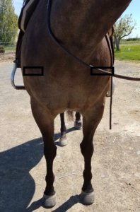
- When viewing the horse from the front, the sound limb drops more in the shoulder. You can put white tape over the point of the shoulder to help see this if needed.
- Watch the fetlock drop. This can be very helpful when there is not an obvious head bob. The fetlock will drop more on the sound limb because the horse is putting more weight on this limb.
- Watch where the foot falls when on a straight line coming toward you. You may be able to see one side of the hoof hit the ground first. Example: the hoof will land on the outside first then the inside.
- You can also try putting two different color wraps or boots on the limbs to help you see the difference in movement.
When a veterinarian evaluates your horse, we do all of these things along with flexion tests. Flexions stress regions of the limb to help the veterinarian narrow down the where the lameness is most likely coming from.
At this point, diagnostic nerve blocks will help us narrow down a region even further. As with the flexion tests, these nerve blocks are not specific to a certain structure (generally), but they get us into the right area.
From here we either take radiographs/x-rays, ultrasound the area, or possibly recommend more advanced imaging like MRI. Once the cause of the lameness is determined, your veterinarian will give different recommendations as to how best to help your horse along with the prognosis.