
When,Why, and How this Diagnostic Tool is Helpful
By James Bryant, DVM, Diplomat ACVS
Our understanding of equine lameness and the availability of new diagnostic tools have increased exponentially over the past 10 years. At the same time, there now is so much new information out there for horse owners to process that certain questions are inevitable.
MRI is the one of the latest tools in our armamentarium for evaluation of the soft tissues and bone of the limb. (Other traditional diagnostic tools for lameness include radiographs, or X-rays, for evaluation of bone and ultrasound for evaluation of soft tissue.) Following are answers to some of the more common questions about equine MRI.
First of all, how does an MRI work?
MRI stands for “magnetic resonance imaging.” An MRI machine uses strong magnetic fields and radiofrequency pulses to produce detailed images, usually of limbs and heads in the case of horses.
MRI technology takes advantage of the natural magnetic properties of hydrogen atoms (water) in the body – creating a very detailed picture of anatomy and physiology. This allows the veterinarian or radiologist to differentiate among soft tissues, bone and body fluids.
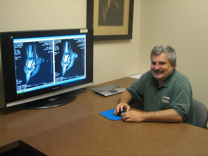
To give its unparalleled look at bone and soft tissue detail, an MRI sections a horse’s limb or other body part into many thin slices – think of a sliced loaf of bread – and in multiple planes (cross section, front to back, etc.). Depending on the tissue that is being examined, typical slice thickness is from 3 mm to 5 mm, with specific scans down to 1 mm.
We can view all the tissues and bone structures of the region from various angles to precisely identify and locate the problem. For most scans, we get images in three planes: sagital, transverse and dorsal, creating a 3-dimensional image of the region.
Currently, there are low-field and high-field MRI systems for equine imaging. The “low” and “high” refer to the magnetic field strength of the system, which determines the resolution of the images and the speed at which the images are acquired. Today, two low-field options exist: standing and recumbent. All high-field MRIs are recumbent.
If your horse’s veterinarian recommends an MRI, discuss the different imaging options with him or her, along with the pros and cons of each MRI system and other diagnostic tools, given your horse’s specific case. At Pilchuck, we offer access to both a low-field recumbent unit and a mobile high-field magnet at the clinic once a month.
Why would my horse need an MRI versus an ultrasound or X-ray?
Used in human medicine for decades, MRIs are gaining traction in equine medicine, especially for orthopedic conditions.
It’s important to note that MRIs don’t replace radiographs or ultrasounds, but are used when those methods do not produce a definitive diagnosis. A thorough lameness evaluation – a critical step in all cases – would include diagnostic nerve blocks to localize the region of injury, then radiographs and ultrasound to look for a cause. If the diagnosis is made from radiographs or ultrasound, then an MRI may not be necessary.
Unlike other imaging modalities, MRI is not a good screening tool due to the limitations of time under anesthesia or sedatives (for standing units); therefore, accurate localization of the lesion with nerve blocks is vital prior to performing an MRI.
What are the best situations to use MRI as a diagnostic tool?
MRI has allowed the visualization of the bone and soft tissue structures in the foot like never before. (The hoof wall can interfere with imaging by other methods, especially ultrasound, but an MRI can get a picture of all those deeper, smaller structures with a super-high level of detail.) In fact, certain injuries can only be picked up with an MRI, such as damage to the cartilage and bone under the cartilage, injuries to the small ligaments and soft tissue structures, and bone bruising.
In addition to evaluation for foot pain, there are many cases in which MRI is an excellent diagnostic tool. In my practice, we routinely use MRIs to diagnose clinical problems in the horse’s foot and pastern, fetlock, metacarpus/metatarsus (high suspensory regions), carpus, tarsus (hock), and head/brain/nasal sinuses.
To go into a little more detail, thanks to MRIs, we veterinarians can now accurately diagnose tears in the deep tendon in the foot, reassess our diagnosis of navicular disease and look at the collateral ligaments of the coffin joint – just to cover a few things. MRIs also allow for the visualization of bone edema (swelling), bone thickening due to trauma, cartilage damage, fractures, and degeneration of bone such as with navicular disease. Simply put, MRIs have proven invaluable in cases where the definitive cause “hides” from traditional imaging tools such as radiographs and ultrasounds.
How will my horse benefit from having an MRI?
MRIs ultimately allow us to be more focused and specific about treatment and follow-up, because we are able to accurately categorize the underlying condition. In my practice, we’ve helped hundreds of horses get diagnoses that wouldn’t be possible without the MRI.
James Bryant, DVM, Diplomat ACVS, is head of Pilchuck Veterinary Hospital’s equine department, where he is a staff surgeon and lameness specialist. After receiving his veterinary degree from the University of California at Davis, College of Veterinary Medicine in 1995, Dr. Bryant completed an internship at Texas A&M University, followed by a surgical residency at the University of Florida.
Published August 2011 Issue
The Northwest Horse Source is an independently owned and operated print and online magazine for horse owners and enthusiasts of all breeds and disciplines in the Pacific Northwest. Our contemporary editorial columns are predominantly written by experts in the region, covering the care, training, keeping and enjoyment of horses, with an eye to the specific concerns in our region.